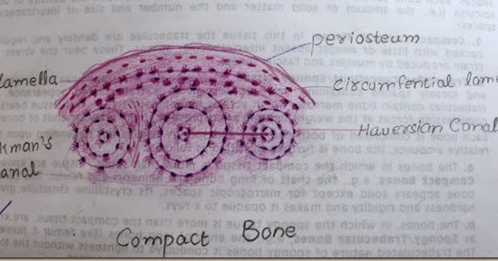
Histology Of Compact Bone Diagram : Bone Histology - Embryology. Compact bone and spongy/cancellous bone are the two types of bones in the human body. This shows the architecture of compact bone which is designed to nourish and regulate osteocytes and bone matrix. The mineral calcium phosphate hardens this framework, giving it strength. (b) enlarged diagram of periosteum and compact bone in (a). Compact bone is dense and composed of osteons, while spongy bone is less dense and made up of trabeculae.
More than 99 percent of our body's calcium is held in our bones and teeth. Labeled diagram of an osteon. Compact bone forms a dense layer on the outside of bones. Given below is a labeled diagram to help you understand the structure of compact long bones, as well as the microscopic structure or histology of the haversian system of compact bones. You may also save it to your computer for more zoomed view.
Histology Slides Database: compact bone high resolution ... from lh3.ggpht.com Compact bone high resolution histology diagram. Osteoblasts deposit the matrix in the form of thin sheets which are called lamellae. Compare and contrast compact and spongy bone. Spongy bone is also referred to as cancellous bone. The objective of the current study. Bone curriculum from the american society for bone and mineral research,. The mineral calcium phosphate hardens this framework, giving it strength. That's a great way for people who are more visual to really see the difference, and it looks cool besides.
Compact bone and spongy bone:
Compact bone high resolution histology diagram. In development there are 2 separate signaling pathways for pattern formation and the formation of bone itself. 5 the standard histology textbook gives the impression that all mammalian bone is fine lamellar bone containing numerous secondary osteones. Compact bone forms a dense layer on the outside of bones. General anatomy and histology of bone. Available at the itunes store(link is external) and for android users at the google play store(link. Start studying histology of compact bone. That's a great way for people who are more visual to really see the difference, and it looks cool besides. Compact bone, microscopically, is made of numerous osteons, whereas spongy bone is composed of sheets of lamellar bone and does not contain osteons. Compare and contrast compact and spongy bone. (b) in this micrograph of the osteon, you can clearly see the concentric. Stability of the compact bone. The mineral calcium phosphate hardens this framework, giving it strength.
In this video lecture we have explained histology of compact bone using high quality histological. Click on the image to enlarge it. Though bone comes in several shapes and sizes the structure and composition of bone is the same in all. This is a first lecture of our new histology series. (b) enlarged diagram of periosteum and compact bone in (a).
HLS [ Cartilage and Bone and Bone Histogenesis, compact ... from www.bu.edu 5 the standard histology textbook gives the impression that all mammalian bone is fine lamellar bone containing numerous secondary osteones. Compare and contrast compact and spongy bone. Histology classification of bone tissue. That's a great way for people who are more visual to really see the difference, and it looks cool besides. The human eye can discern only two types of bone. In simple terms bone is a type of specialised connective tissue. Feel free to use for study purposes. The mineral calcium phosphate hardens this framework, giving it strength.
This is a first lecture of our new histology series.
Spongy bone is also referred to as cancellous bone. Compact bone high resolution histology diagram. Bones have an internal structure similar to a honeycomb, which makes. This article about bones explains the fundamentals of anatomy for physicians. In simple terms bone is a type of specialised connective tissue. Compare and contrast compact and spongy bone. General anatomy and histology of bone. In development there are 2 separate signaling pathways for pattern formation and the formation of bone itself. A bone is a rigid tissue that constitutes part of the vertebrate skeleton in animals. The hollow region in the diaphysis is called the medullary cavity, which is filled with yellow marrow. Bone also serves as a reservoir of calcium, phosphate, and other ions that can be released or stored in a controlled fashion to maintain constant in addition, bones form a system of levers that multiply the forces generated during skeletal muscle contraction and transform them into bodily movements. Cortical bone is compact bone, while cancellous bone is trabecular and spongy bone. Bones are mostly made of the protein collagen, which forms a soft framework.
Compare and contrast compact and spongy bone. Histology classification of bone tissue. Start studying histology of compact bone. This shows the architecture of compact bone which is designed to nourish and regulate osteocytes and bone matrix. More than 99 percent of our body's calcium is held in our bones and teeth.
Structure and Function of the Haversian System Explained ... from i.pinimg.com Compact bone and spongy/cancellous bone are the two types of bones in the human body. Labeled diagram of an osteon. In mature compact bone most of the individual lamellae form concentric rings around larger longitudinal canals (approx. Cortical bone is compact bone, while cancellous bone is trabecular and spongy bone. It is found beneath the periosteum of all bones and makes up the bulk of the diaphyses of long bones. For a surgeon this distinction is what he encounters in the operating room. Given below is a labeled diagram to help you understand the structure of compact long bones, as well as the microscopic structure or histology of the haversian system of compact bones. For exams it is more important for physicians to understand the structure and composition of image:
Compact bone and spongy/cancellous bone are the two types of bones in the human body.
(b) in this micrograph of the osteon, you can clearly see the concentric. In this video lecture we have explained histology of compact bone using high quality histological. The strongest form of bone tissue. Bones have an internal structure similar to a honeycomb, which makes. The mineralized tissue is seen as spicules. Histology classification of bone tissue. Blood vessels and nerves enter the bone through the nutrient foramina to nourish and innervate bones. Though bone comes in several shapes and sizes the structure and composition of bone is the same in all. The objective of the current study. In development there are 2 separate signaling pathways for pattern formation and the formation of bone itself. A bone is a rigid tissue that constitutes part of the vertebrate skeleton in animals. Compact bone forms the outer 'shell' of bone. Compact bone is dense and composed of osteons, while spongy bone is less dense and made up of trabeculae.
Blood vessels and nerves enter the bone through the nutrient foramina to nourish and innervate bones compact bone diagram. That's a great way for people who are more visual to really see the difference, and it looks cool besides.
Share :
Post a Comment
for "Histology Of Compact Bone Diagram : Bone Histology - Embryology"


Post a Comment for "Histology Of Compact Bone Diagram : Bone Histology - Embryology"