Shoulder Ligament Anatomy Diagram - Anatomy Lesson Shoulder Musculature Beautiful To The Core Shoulder Muscle Anatomy Muscle Anatomy Shoulder Anatomy. Bones of the upper limb. Robin smithuis and henk jan van der woude. Human anatomy diagrams show internal organs, cells, systems, conditions, symptoms and sickness information and/or tips for healthy living. Shoulder separation describes the condition in which the ligaments connecting the ac joint are injured and the acromion begins to move away from the clavicle. The most common shoulder injuries involve the muscles, ligaments, cartilage, and tendons, rather than the bones.
The shoulder joint, also known as the glenohumeral joint, is a ball and socket joint with the most extensive range of motion in the human body. Several ligaments make up parts of the joint capsule, and these ligaments are important in keeping the shoulder joint in proper position. This diagram depicts anatomy glenohumeral joint shoulder ligaments en medical. Anatomy, movement & muscle involvement » how to relief. Pectoral girdle and shoulder anatomy tutorials.
Shoulder Anatomy Springerlink from media.springernature.com Last update september 3, 2020. All about the shoulder muscles. This webmd article explains what and where ligaments are and how you can injure ligaments are bands of tough elastic tissue around your joints. Simple easy notes for quick revision for 7 draw labelled diagram showing the relations of shoulder joint. Instant anatomy is a specialised web site for you to learn all about human anatomy of the body with diagrams, podcasts and revision questions. Divided into two additional ligaments including the trapezoid ligament. Various types of injuries and degenerative conditions can cause the shoulder to become painful. Sechrest, md narrates an animated tutorial on the basic anatomy of the shoulder.
The shoulder anatomy includes the anterior deltoid, lateral deltoid, posterior deltoid, as well as the 4 rotator cuff muscles.
Anatomy, movement & muscle involvement » how to relief. Moore clinically oriented anatomy, (7th ed.). Check out our articles, video tutorials, quizzes, and labeled diagrams. This diagram depicts anatomy glenohumeral joint shoulder ligaments en medical. Several ligaments make up parts of the joint capsule, and these ligaments are important in keeping the shoulder joint in proper position. Robin smithuis and henk jan van der woude. The shoulder joint is the connection between the chest and the upper extremity. Joints, ligaments and connective tissues. Slap (superior labrum anterior to posterior) tear. 259) may become ossified, transforming the scapular notch into an anomalous bony canal, the scapular foramen. This webmd article explains what and where ligaments are and how you can injure ligaments are bands of tough elastic tissue around your joints. The pectoral girdle or shoulder girdle consists of four bones that connect these supporting bones to the trunk. Want to learn more about the types of body movements?
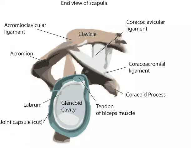
Shoulder separation describes the condition in which the ligaments connecting the ac joint are injured and the acromion begins to move away from the clavicle. Bones of the upper limb. Notice superior labrum and attachment of the superior glenohumeral ligament. Your shoulder is made up of three bones: Diagram of the human shoulder joint, back view.
Common Injuries Of The Shoulder Online Medical Encyclopedia Shoulder Anatomy Shoulder Bones Arm Bones from i.pinimg.com The shoulder joint (glenohumeral joint) is a ball and socket joint between the scapula and the humerus. The shoulder joint is the connection between the chest and the upper extremity. 8 name the arteries and the nerves that supply shoulder joint. Sechrest, md narrates an animated tutorial on the basic anatomy of the shoulder. Shoulder bones and ligaments anatomy. They connect bone to bone, give your joints support, and limit their movement. Movements of the human shoulder represent the result of a complex dynamic interplay of structural bony anatomy and biomechanics, static ligamentous and tendinous restraints, and dynamic muscle forces. Last update september 3, 2020.
Shoulder stability is achieved through the interplay of both static and dynamic stabilisers, which work in synchrony to maintain shoulder.
All about the shoulder muscles. Normal anatomy, variants and checklist. Anatomy, movement & muscle involvement » how to relief. Various types of injuries and degenerative conditions can cause the shoulder to become painful. Ligaments of the shoulder joint (hansen, 2009, pg. The shoulder joint is the connection between the chest and the upper extremity. Because of its location superior to the glenohumeral joint, it acts as a protection to the joint. Home > blog > anatomy > shoulder anatomy: Notice superior labrum and attachment of the superior glenohumeral ligament. Human anatomy diagrams show internal organs, cells, systems, conditions, symptoms and sickness information and/or tips for healthy living. This webmd article explains what and where ligaments are and how you can injure ligaments are bands of tough elastic tissue around your joints. This diagram depicts anatomy glenohumeral joint shoulder ligaments en medical. In this episode of eorthopodtv, orthopaedic surgeon randale c.
Diagram of the human shoulder joint, back view. Once stretched, they tend to stay stretched there are several important ligaments about the shoulder girdle. Shoulder bones and ligaments anatomy. Shoulder stability is achieved through the interplay of both static and dynamic stabilisers, which work in synchrony to maintain shoulder. Your shoulder is made up of three bones:
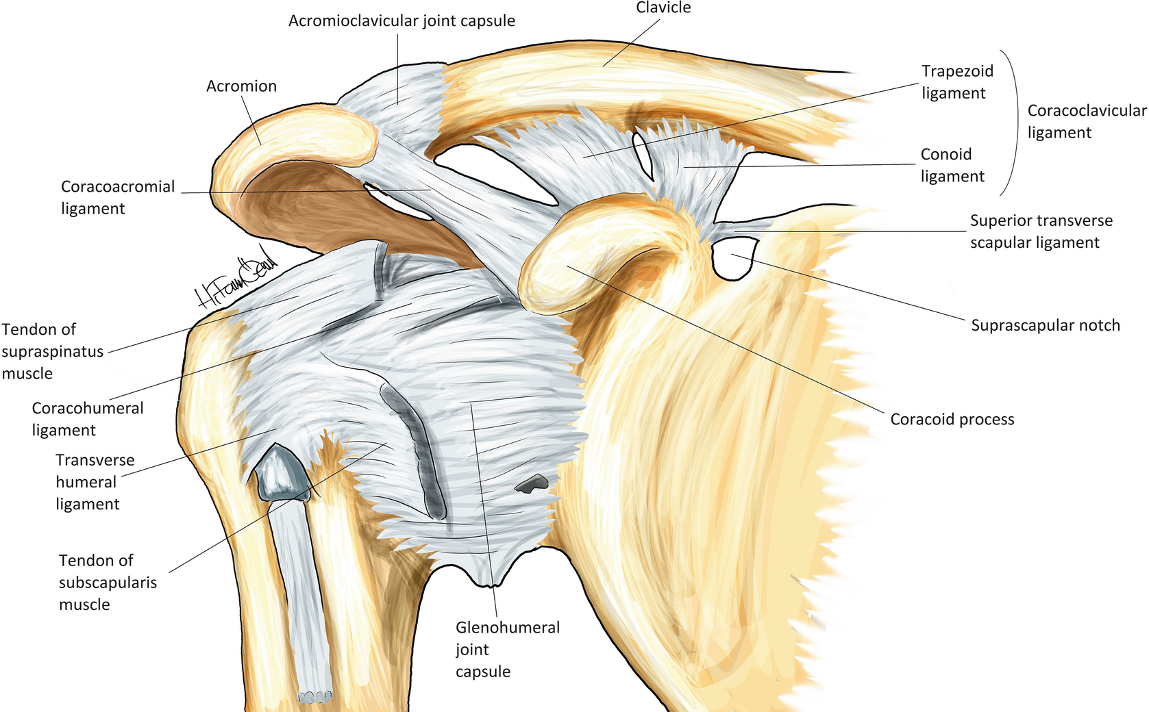
The Anatomy Of The Shoulder from www.ortho.wustl.edu They connect bone to bone, give your joints support, and limit their movement. This webmd article explains what and where ligaments are and how you can injure ligaments are bands of tough elastic tissue around your joints. Learn about shoulder anatomy and watch anatomy of the shoulder video's presented by joi. This diagram depicts anatomy glenohumeral joint shoulder ligaments en medical. The coracohumeral ligament originates on the base and outside the border of the coracoid process of the scapula and attaches to a landmark on the humerus called the greater tuberosity. Instant anatomy is a specialised web site for you to learn all about human anatomy of the body with diagrams, podcasts and revision questions. Shoulder stability is achieved through the interplay of both static and dynamic stabilisers, which work in synchrony to maintain shoulder. Notice superior labrum and attachment of the superior glenohumeral ligament.
The shoulder joint (glenohumeral joint) is a ball and socket joint between the scapula and the humerus.
Ligament, tough fibrous band of connective tissue that serves to support the internal organs and hold bones together in proper articulation at the joints. Because of its location superior to the glenohumeral joint, it acts as a protection to the joint. Human anatomy diagrams show internal organs, cells, systems, conditions, symptoms and sickness information and/or tips for healthy living. Bones of the upper limb. Your upper arm bone (humerus), your shoulder blade (scapula), and your collarbone (clavicle). 8 name the arteries and the nerves that supply shoulder joint. Diagram of the human shoulder joint, back view. Learn all about the anatomy and function of the shoulder girdle fast and efficiently in this article, where we outline its muscles, bones, joints and more. Last update september 3, 2020. The transverse humeral ligament is not shown on this diagram/caption. The shoulder joint is the connection between the chest and the upper extremity. The shoulder is a complex combination of bones and joints where many muscles act to provide the widest range of motion of any part of the body. Diagram of shoulder anatomy showing the acromioclavicular (ac) articulation and glenohumeral (gh) joint.
Home > blog > anatomy > shoulder anatomy: shoulder anatomy diagram. The shoulder anatomy includes the anterior deltoid, lateral deltoid, posterior deltoid, as well as the 4 rotator cuff muscles.
Share :
Post a Comment
for "Shoulder Ligament Anatomy Diagram - Anatomy Lesson Shoulder Musculature Beautiful To The Core Shoulder Muscle Anatomy Muscle Anatomy Shoulder Anatomy"


Post a Comment for "Shoulder Ligament Anatomy Diagram - Anatomy Lesson Shoulder Musculature Beautiful To The Core Shoulder Muscle Anatomy Muscle Anatomy Shoulder Anatomy"